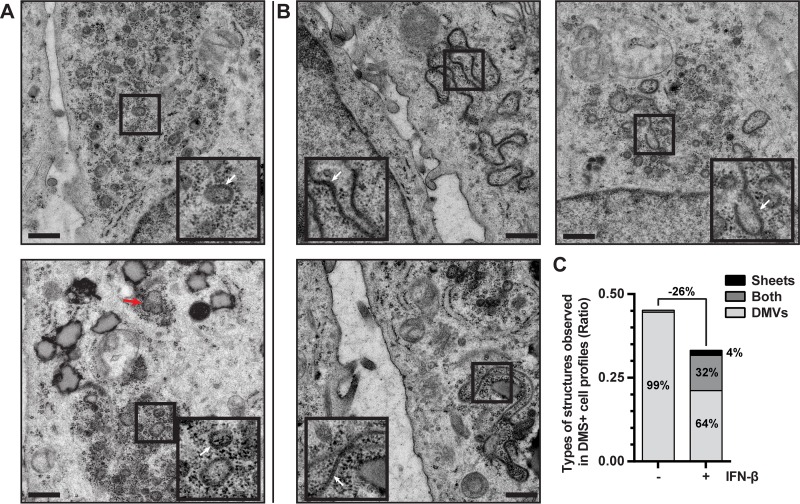
FIG 4 .
IFN-β treatment blocks DMV formation and leads to the accumulation of double-membrane sheets. (A and B) EM images of HuH-7/tetR/HA-nsp2-3GFP cells in which expression is induced with tetracycline for 24 h. Cells represented in images of panel B were simultaneously treated with 500 U/ml IFN-β for 24 h. Images were extracted from mosaic maps used for quantifications, and scale bars represent 500 nm. Insets are 2× magnifications of areas where DMVs (A) or double-membrane sheets (B) are visible (indicated with white arrows). The different appearance of these samples relative to the data shown in Fig. 2 is the result of the different sample preparation approaches (chemical fixation versus HPF-FS, respectively). (C) The occurrence of the different types of DMS (DMVs, double-membrane sheets, or both) in the cell profiles is shown in control or IFN-β-treated HuH-7/tetR/HA-nsp2-3GFP cells that were tetracycline induced.

