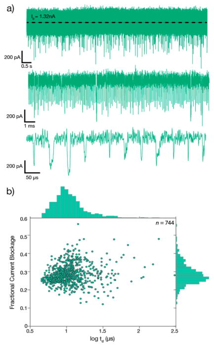
Figure 5.
Single-stranded DNA transport through a 2.3 nm diameter MoS2 nanopore. (a) Three-second continuous current trace for a 2.3 nm diameter pore after the addition of 20 nM 153-mer ssDNA to the cis chamber ([KCl] = 0.40 M, Vtrans = 200 mV, sampling rate =4.17 MHz, data low-pass filtered to 200 kHz). Concatenated sets of events at different magnifications are shown below the trace. (b) Scatter plot of fractional current blockade (see text) vs dwell time td, as well as histograms of each parameter shown in each corresponding axis (n = number of molecules detected).

