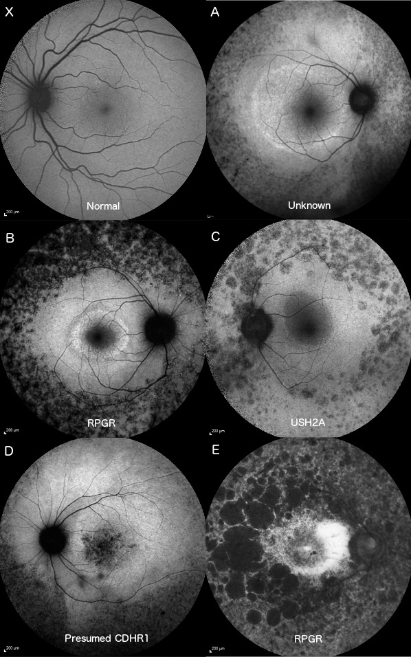
Figure 3.

Fifty-five–degree AF images of a normal patient (X) and the control eye of the five participants. Participants A–C had recognizable parafoveal rings of hyperfluorescence with granular peripheral hypofluorescence. Participant D had some additional granular foveal hyperfluorescence and a sectoral peripheral hypofluorescence. Participant E retained a poorly defined parafoveal arc with large, scalloped peripheral regions of hypofluorescence.
