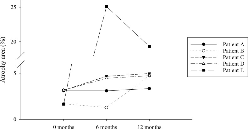
Figure 5.
Changes in area of the AF image under the 10th percentile, chosen as a surrogate for measurement of RPE and photoreceptor atrophy. Participants A–D exhibited an increase in peripheral hypofluorescence. The advanced disease of participant E led to highly fluctuant results suggesting that the method is not as reliable in patients with late disease.

