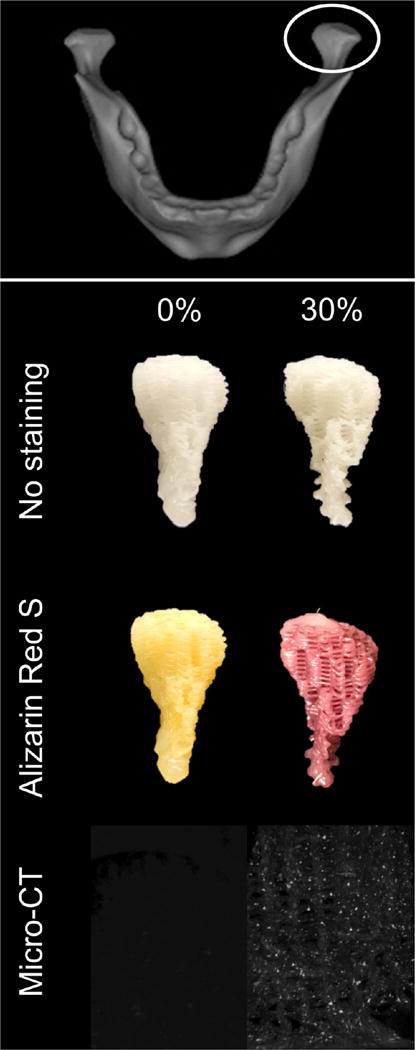
Figure 9.

Top: Anatomical shape printing of pure and hybrid scaffolds. Middle: A human temporomandibular joint condyle was isolated and printed into anatomically shaped, porous scaffolds. Scaffolds were subject to Alizarin Red S staining to confirm and visualize the presence of mineralized particles in the hybrid scaffold. Bottom: MicroCT scans performed to confirm the presence of mineralized particles in the 30% DCB:PCL scaffolds. There were no mineral particles in the pure PCL scaffold.
