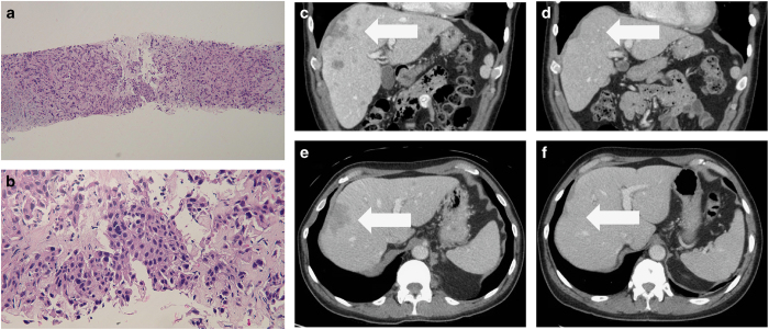
Figure 1.
Histology of metastatic basal cell Carcinoma sample—medium (a) and high (b) magnification views of liver biopsy and CT scans demonstrating near complete response to PD-1 blockade (e, f). The metastatic tumour extensively infiltrates the liver; several tumour cells have large pleomorphic nuclei (a). Aggregates of the basaloid tumour cells are also present (b). (Hematoxylin and eosin: ; ×10, a; ×40, b). Baseline CT of liver lesions before treatment (c, e). Near complete resolution of the liver lesion after nivolumab for four months (d, f). The white arrows demonstrate metastatic basal cell carcinoma in the liver prior to (c, e) and following (d, f) 4 months of treatment with nivolumab.

