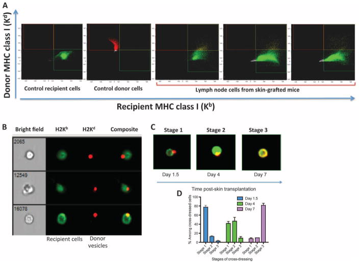
Fig. 2. Detection of donor leukocytes in LNs of skin-grafted mice.
LN cells (ipsilateral axillary and brachial LNs) from naïve B6 and BALB/c mice as well as B6 mice recipient of a BALB/c skin allograft were collected at different time points after transplantation. The cells were stained with anti-MHC class I Kb antibodies and anti-MHC class I Kd bound to FITC and allophycocyanin, respectively. The presence of recipient (Kb+) and donor (Kd+) cells was assessed using flow imaging with low-speed fluidics at ×40 magnification. Data analysis was performed using IDEAS 6.1 image processing and statistical analysis software (Amnis, EMD Millipore). (A) Plots obtained with control recipient cells, control donor cells, and cells from three different transplanted mice tested at day 2 after transplantation. The results are representative of eight mice per group tested in two separate experiments. (B) Representative microscopic analysis of double-positive cells (Kb+/Kd+) observed in (A). The bright field represents the actual optical image of the cells (×40 magnification). The other columns show the fluorescence of cells. (C) Representative images of double-positive cells (stages 1 to 3) obtained at different time points after transplantation. Stage 3 corresponds to recipient leukocytes cross-dressed with donor MHC. (D) Percentages of recipient leukocytes cross-dressed with donor MHC (stage 3) among double-positive cells found in LNs of skin-grafted mice examined individually at different time points after transplantation ± SEM. The results are representative of six mice tested individually at each time point. P values using unpaired Student’s t test: stage 1: d1.5 versus d4, P = 0.0001; d1.5 versus d7, P = 0.0001; d4 versus d7, P = 0.0022; stage 2: d1.5 versus d4, P = 0.0001; d1.5 versus d7, P = 0.0051; d4 versus d7, P = 0.0001; stage 3: d1.5 versus d4, P = 0.1701 [not significant (NS)]; d1.5 versus d7, P = 0.0001; d4 versus d7, P = 0.0001.

