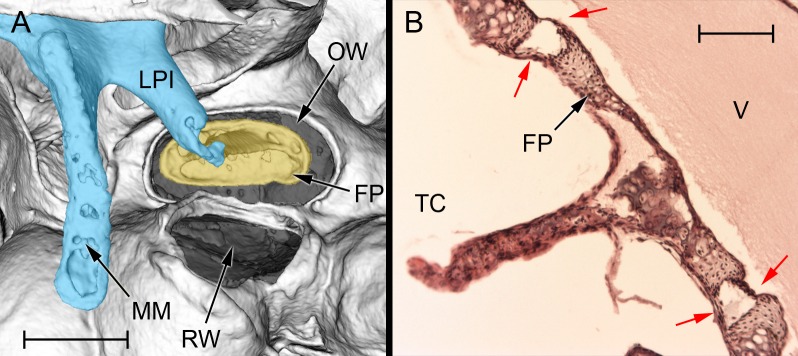
Fig 4. The stapes of Heterocephalus, in situ.
A: MicroView reconstruction of the left stapes region in an adult mole-rat (65 months), ventrolateral view. The malleoincudal complex has been coloured blue, the stapes yellow. Note the very wide gap between stapes footplate and the rim of the oval window. This gap and the opening of the round window (both shaded grey) are covered over by membranes, but these do not show up on the CT reconstruction. Scale bar 0.5 mm. B: Histological section of the stapes of a neonatal Heterocephalus, showing the synovial structure of the stapedio-vestibular articulation. The red arrows indicate the synovial joint capsule, which lies between the stapes footplate and the rim of the oval window. Scale bar 0.1 mm. APSC = ampulla for posterior semicircular canal; ASC = anterior semicircular canal; C = cochlea; DMC = dorsal mastoid cavity; ED = bony tube for endolymphatic duct; ER = epitympanic recess; FP = footplate of stapes; HM = head of malleus; LA = lenticular apophysis of incus; LPI = long process of incus; LSC = lateral semicircular canal; MI = malleoincus; MM = manubrium of malleus; OW = oval window; PC = posterior crus of stapes; PD = bony tube for perilymphatic duct (canaliculus cochleae); PMC = posteromedial mastoid cavity; PSC = posterior semicircular canal; RW = round window; SPI = short process of incus; TC = tympanic cavity; TM = tympanic membrane; TT = tensor tympani tendon; V = vestibule of inner ear; VMC = ventral mastoid cavity.

