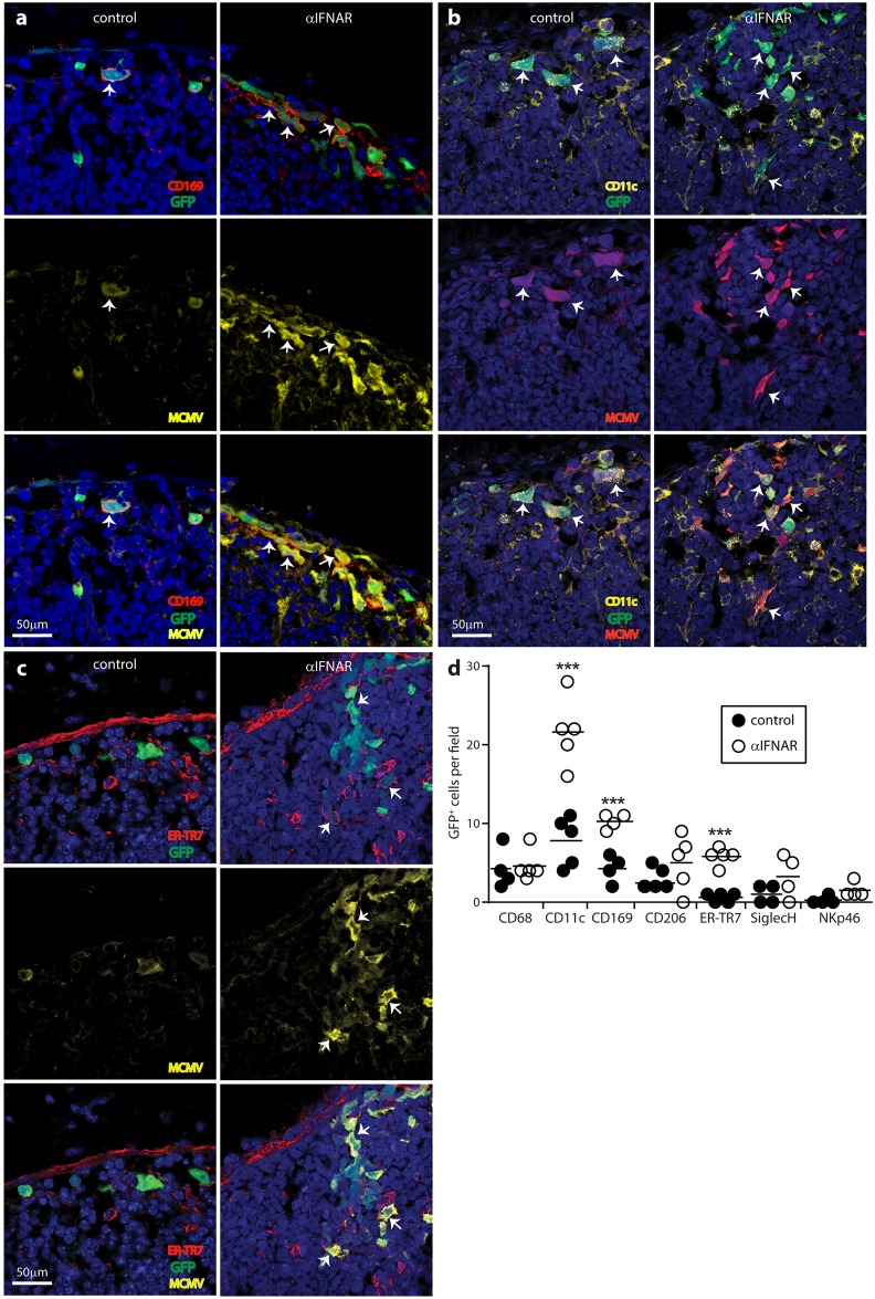
Fig 3. IFNAR blockade increases SSM and FRC infections.
C57BL/6 mice were given IFNAR blocking antibody (αIFNAR) or not (control), then MCMV-GR i.f. (106 p.f.u.). PLN taken 1 day later were stained for viral GFP (GFP) and lytic antigens (MCMV), plus the SSM marker CD169 (a), the SSM/DC marker CD11c (b) or the FRC marker ER-TR7 (c). Nuclei were stained with Hoechst 33342 (blue). Arrows show example infected SSM and FRC. Both were more numerous after αIFNAR. In (d) we counted cells across 4–5 fields of view for PLN sections from each of 3 mice per group. Circles show individual means, bars show group means. Significant differences are indicated (Student’s two tailed unpaired t-test; ***, P<0.001; ****, P<0.0001). We quantified infection also for CD68+ (pan-macrophage/DC), CD206+ (mannose receptor, absent from SSM), SiglecH+ (pDC) and NKp46+ (NK) cells.

