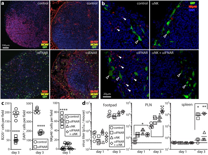
Fig 5. IFN-I-independent NK cell recruitment aids infection control.
(a). C57BL/6 mice were given IFNAR blocking antibody (αIFNAR) or not (control) then i.f. MCMV-GR (106 p.f.u.). 3 days later PLN were stained for viral GFP, lytic antigens (MCMV), and B cells (B220) or FRC (ER-TR7). Nuclei were stained with DAPI. αIFNAR reduced B220 and DAPI staining, and disorganized ER-TR7 staining, implying loss of LN cellularity and structure. (b). C57BL/6 mice were given αIFNAR, depleted of NK1.1+ cells with mAb PK136 (αNK), given both treatments, or given neither (control). All were then given i.f. MCMV-GR (106 p.f.u.). 1 day later PLN were stained for viral GFP and NKp46+ NK cells. Nuclei were stained with DAPI. Closed arrows show example NKp46+ cells. Open arrows show example GFP+ infected cells. (c). Quantitation of staining as illustrated in (a) and (b), across 7 fields of view for sections from each of 5 mice per group, showing significant loss of LN cellularity in IFNAR treated-mice (B220+, DAPI+) and significant NK cell recruitment (NKp46+) after αIFNAR and effective NK cell depletion by mAb PK136. Bars show group means, other symbols show mean counts of individual mice. (****, P<0.0001; Student’s two-tailed unpaired t-test). (d). Organs of mice treated as in (b) were plaque assayed for infectious virus. Bars show means, other symbols show individual mice. Significant differences are indicated (Student’s two tailed unpaired t-test; *p<0.05, **p<0.01). Dotted lines show assay sensitivity limits.

