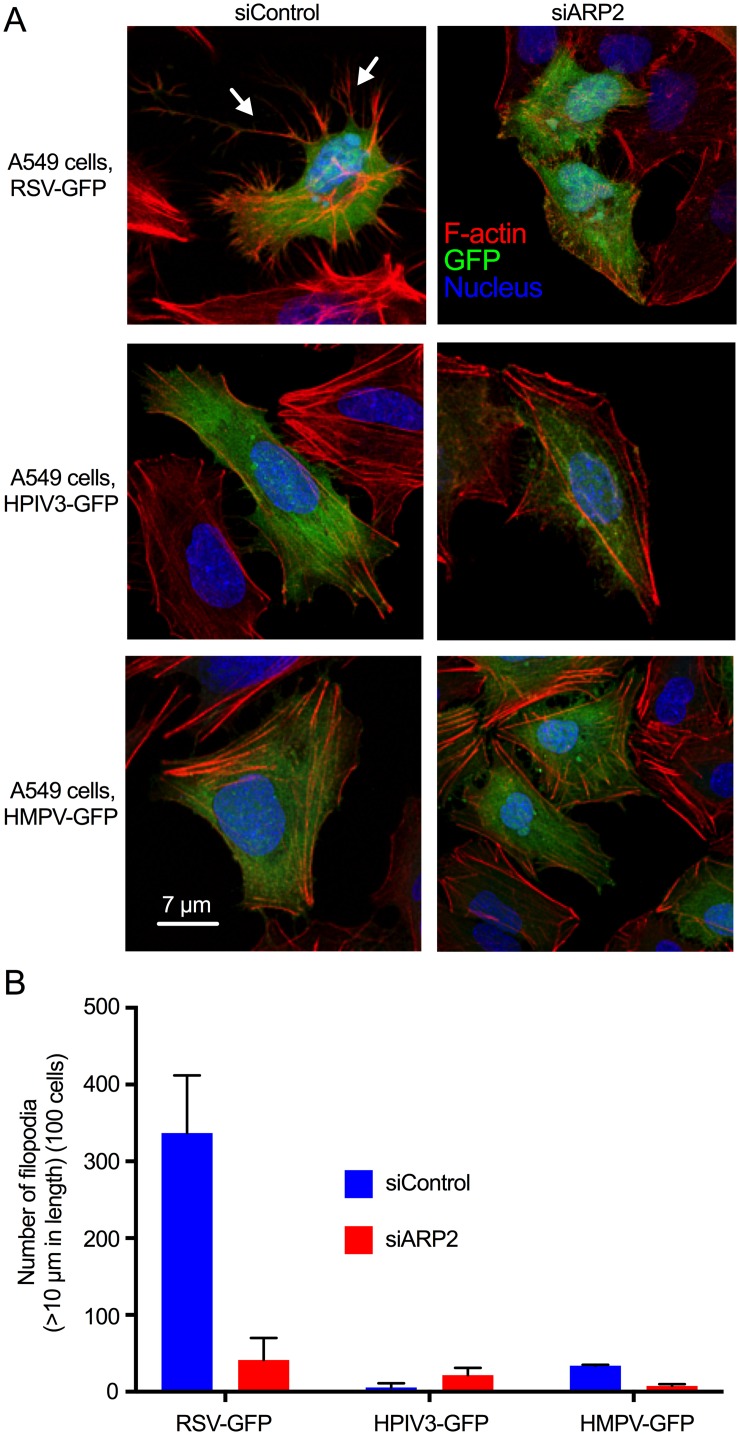
Fig 10. RSV induced filopodia more robustly than HPIV3 or HMPV infection.
(A) A549 cells were transfected with siARP2 or siControl. 48 hr after transfection, cells were infected with RSV-GFP, HPIV3-GFP or HMPV-GFP at MOI = 1. At 24 hpi, cells were fixed and permeabilized, and F-actin was stained with rhodamine phalloidin. Nuclei were stained with DAPI. Infected cells were detected by GFP expression. RSV-induced filopodia are indicated with arrows. (B) The number and length of filopodia were evaluated by automated scanning using confocal microscopy. In brief, Z-stacking for GFP for virus, DAPI for nuclei, rhodamine phalloidin for F-actin was performed for 50 to 100 different random fields of interest in each coverslip. The length and number of filopodia was measured on 100 cells per treatment per experiment from the surface to the tip of the filopodium. Data were combined from two independent experiments. Error bar: SD.

