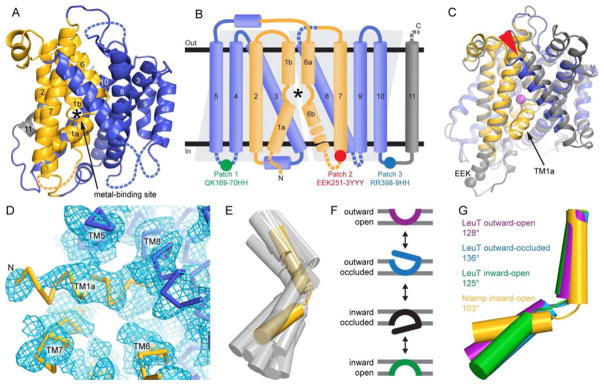
Figure 1.
DraNramp structure in the inward-facing state shows a highly kinked TM1. (A) Cartoon representation of DraNramp with TM helices labeled; the bundle (TMs 1, 2, 6, and 7) is gold, scaffold (TMs 3, 4, 5, 8, 9, and 10) blue, and TM11 gray. Dashed loops are disordered in the structure. (B) DraNramp topology diagram, with helices as cylinders, and gray trapezoids highlighting the inverted structural repeats (TMs 1-5 and TMs 6-10). Intracellular loop mutations Patch 1 to 3 in the crystallized construct are indicated. (C) Superposition of DraNramp (blue and gold) and ScaNramp (4WGW; gray and Mn2+ magenta) indicates a similar overall fold. The main differences are the position of TM5 (red arrowhead) and the presence of TM1a in DraNramp. Grey spheres mark the ScaNramp EEK motif corresponding to Patch 2, disordered in DraNramp. (D) Final 2Fo-Fc electron density map at 0.8σ showing density for TM1a. DraNramp is represented as a Cα trace. (E) Comparison of TM1 kink angle of DraNramp (yellow) with published LeuT-fold structures (gray; LeuT 2A65, 3TT1, 3TT3, 5JAE; Mhp1 2JLN, 4D1B, 2×79; vSGLT 2XQ2, 3DH4; BetP 4LLH, 4AIN, 4DOJ, 4C7R). The scaffolds of the corresponding structures were superimposed and oriented as in (A). (F) Conformational states in a transport cycle, color-coded as in (G). (G) The TM1b helices were aligned for DraNramp and three distinct LeuT conformations, highlighting the common kink at the unwound substrate-binding region in the middle of TM1. The kink is even more pronounced in inward-open DraNramp than any LeuT structure. See also Figure S1.

