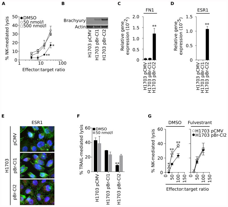
Figure 3. Fulvestrant renders mesenchymal cells more sensitive to immune-mediated lysis.
(A) Susceptibility of parental H460 cells treated with two doses of fulvestrant vs. DMSO to lysis by NK cells at various effector-to-target ratios. (B) Brachyury protein levels and expression of mRNA encoding for fibronectin (C) and ESR1 (D), relative to GAPDH, in clonally-derived H1703 cells transfected with pCMV vs. pBr (Clones 1 and 2). (E) Immunofluorescent analysis of ESR1 (pink signal) protein expression in H1703 pCMV, pBr-Cl1 and pBr-Cl2 cells (100× magnification). Green and blue correspond to phalloidin and DAPI staining, respectively. (F) Susceptibility to TRAIL-mediated lysis in cells pre-treated with fulvestrant (grey bars) vs. DMSO (black bars). (G). Susceptibility of H1703 pCMV and H1703 pBr-Cl2 cells treated with fulvestrant (right panel) vs. DMSO (left panel) to lysis by NK cells at various effector-to-target ratios. Error bars indicate the standard error of the mean (SEM) of triplicate measurements. [* p<0.05, ** p<0.01].

