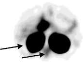
Table 1.
Initial findings with reconstruction settings of clinical scans
| Flash 3D | Hybrid Recon | |
|---|---|---|
| Version | Siemens Syngo MI.SPECT application E.soft 2009a, 8.1.15.7 service pack 2 | HERMES P5 GOLD 4.6A Hybrid Recon 1.1.2 |
| Reconstruction algorithm | OSEM3D | OSEM3D, MAPMRP and MAPsmoothing |
| # iterations–# subsets | 6–16 | 4–16 |
| Attenuation correction | CT-based | CT-based |
| Scatter correction | Triple energy window, SPECT-based | CT, Monte Carlo-based |
| Both photopeaks of 111In reconstructed | Yes | Yes |
| Collimator correction | Mathematically calculated 3D cone beam modelling | Mathematically calculated 3D cone beam modelling |
| Post reconstruction filtering | Gaussian FWHM = 0.84 cm | Gaussian FWHM = 0.96 cm |
| Most obvious artefacts in the initial human images | Large low-intensity rim around kidneys 
|
Small low-intensity rim around the kidneys, somewhat increased intensity in the vertebrae area 
|
