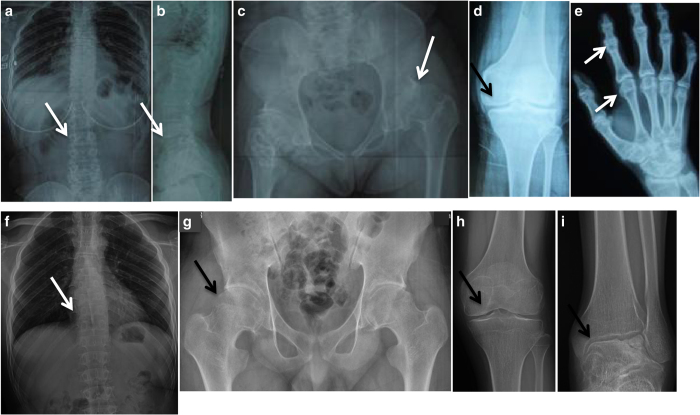
Figure 1.
Radiographic features of the two patients with progressive pseudorheumatoid dysplasia. (a–e) Patient 1 at age 15. (f–i) Patient 2 at age 17. (a, b) Thoracolumbar spine. Platyspondyly with anterior breaking of the vertebral bodies. (c) Pelvis. Narrow joint spaces and subluxation of the hip joints and irregular densities and cyst-like structures in the femoral head and neck. (d) Knee. The articular surface was flat and irregular, and the joint space was narrow. (e) Hand. Enlargement of the interphalangeal and metacarpal epiphyses. (f) Chest. Decreased heights of the vertebrae. (g–i) Large joints of the lower extremities showed narrow joint spaces.

