Figure 2.
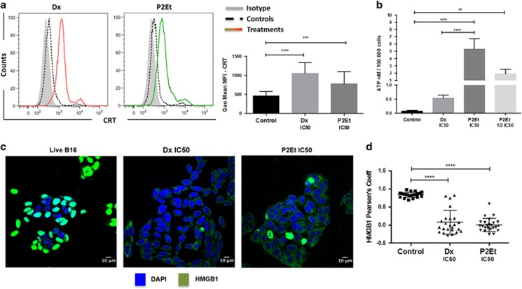
P2Et fraction induces immunogenic B16F10 cell death. B16F10 melanoma cells were cultured in complete media and treated with P2Et IC50 (63.5 μl/ml), Dx IC50 (0.06 μg/ml) or with the negatives controls (EtOH and DMSO—vehicles), for 24 h in all cases. (a) CRT: the surface exposure of CRT was determined by flow cytometry among viable cells (Aqua vivid negative), comparisons between negative controls (dashed line—black), positive control Dx (red line) and P2Et-treated cells (green line) and a isotype control (solid histogram). Bars represent the geometric mean values±S.E.M. (n=5), ***P<0.001; ****P<0.0001. (b) ATP: cells were maintained in control conditions or treated with P2Et IC50, Dx IC50, followed by the assessment of ATP secretion in culture supernatants by luminescence. Quantitative data are means±S.E.M. (n=3). **P<0.01; ***P<0.001; ****P<0.0001. (c) HMGB1: mobilization was determined by confocal microscopy. Images were acquired with Olympus FV1000 with a 60 × PlanAPO objective and are representative of three independent experiments. Primary antibody for HMGB1 (rabbit anti-mouse) was detected using conjugate goat anti-rabbit secondary antibody (Alexa Fluor 488—green) and nuclei stained with DAPI. (d) Pearson correlation coefficients: the distribution coefficients for HMGB1, each dot is one cell for a total of 50 cells per group, values are means±S.E.M. (n=3) **** P<0.0001

