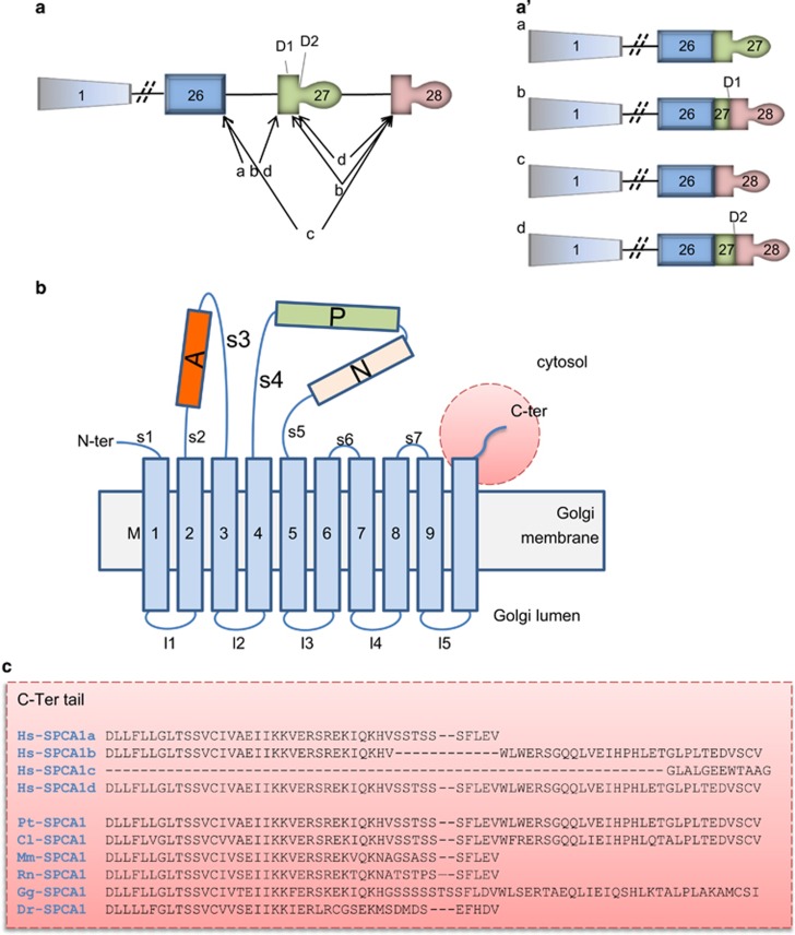
Figure 1.
Representation of the ATP2C1 gene alternatively spliced and the molecular structure of encoded SPCA1. (a) The ATP2C1 gene consists of twenty-eight exons (represented by boxes), which are alternatively spliced as indicate by the internal 5′ donor splice sites, D1 and D2 generating four different mRNA. Diagonal lines illustrate the slicing patterns generating splice variants ATP2C1a-d. (a′) The ATP2C1a-d splice variants are schematically represented. (a) and (a′) are modified from Micaroni and Malquori.50 (b) Actuator domain (A), phosphorylation domain (P), nucleotide-binding domain (N) and 5 stalk helices (S) in the cytoplasm, and 10 transmembrane helices (M). This figure was adapted from Matsuda et al.8 (c) In gray is the exon 26, in yellow the exon 27, in green the exon 28. According to the present literature, the isoform SPCA1c seems not be coded in a protein. This isoform is missing the exon 27 coding for the transmembrane 10 (M10). Furthermore, this isoform is missing the possibility to have a cytosolic C-terminal tail where potential binding sites for other proteins is present, reinforcing the idea that this isoform is not functional. Hs-SPCA1a (NP_055197); Hs-SPCA1b (NP_001001487.1); Hs-SPCA1c (NP_001001485.2); Hs-SPCA1d (NP_001001486.1); Pt-SPCA1 (XP_001145788.1); Cl-SPCA1 (XP_534262.2); Mm-SPCA1 (NP_778190.3); Rn-SPCA1 (NP_571982.2); Gg-SPCA1 (XP_015137243.1); Dr-SPCA1 (XP_003200287). Hs: Homo sapiens; Pt: Pan troglodytes; Cl: Canis lupus; Mm: Mus musculus; Rt: Rattus norvegicus; Gg: Gallus gallus; Dr: Danio rerio

