Figure 8.
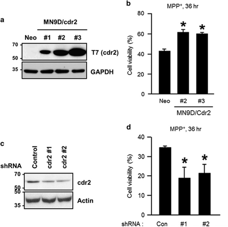
Protective role for cdr2 in MPP+-induced cell death. (a) MN9D cells were stably transfected with T7-tagged mouse cdr2 (MN9D/cdr2 #1, #2, or #3) or empty vector (MN9D/Neo). Expression levels were validated by immunoblot analysis using mouse monoclonal anti-T7 antibody. (b) MN9D/Neo and two highly expressing MN9D/Cdr2 cell lines were treated with 50 μM MPP+ for 36 h. Cell viability was measured using MTT reduction assay and expressed as a percentage of that for untreated control cells. Data are shown as the mean±S.D. of three independent experiments. *P<0.05. (c) MN9D cells were stably transfected with cdr2 shRNA- or control shRNA-expressing vectors. Extent of cdr2 knockdown was determined by immunoblot analysis using anti-cdr2 antibody. (d) After treatment with 50 μM MPP+ for 36 h, MTT reduction assay was performed. Cell viability was expressed as a percentage of that for untreated control cells. Data are shown as the mean±S.D. from three independent experiments. *P<0.05

