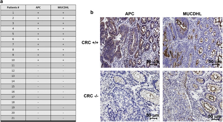
Figure 1.
(a) Table summarizing the results of the immuno-histochemical analysis of APC and μ-protocadherin (MUCDHL) protein levels in 21 CRC samples; + and − indicate presence or absence of the analyzed protein, respectively. (b) Immuno-histochemical analysis of two representative CRC cases exhibiting a double positive (APC+/MUCDHL+) (CRC +/+) or a double negative (APC−/MUCDHL−) (CRC −/−) expression pattern. Positivity appears as a dark staining; cell nuclei were counterstained with hematoxylin

