Figure 1.
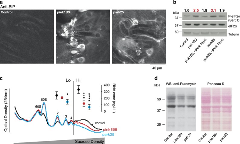
Activation of phospho-eIF2α signalling and attenuation of translation in pink1 and parkin mutant flies. (a) Increased levels of BiP in the body wall muscle of pink1 and parkin mutant larvae. Representative confocal images with the indicated genotype stained with α-BiP antibody are shown. (b) Increased levels of phospho-eIF2α in pink1 and parkin mutant flies are reduced by knockdown of dPerk. Whole-fly lysates were analysed using the indicated antibodies. Ratios of signal intensity between phospho and total-eIF2α are shown at the top. (c) Polysomal distribution of mRNAs of young adult male flies showing individual ribosomal subunits and the polysome peaks. RNA concentrations were measured from the low (Lo) and high (Hi) translation fractions (mean±S.D., asterisks, one-way ANOVA with Dunnett's multiple comparison test; n=4). (d) Reduced puromycin incorporation in nascent proteins in pink1 and parkin mutant flies. Whole-fly lysates were analysed with an anti-puromycin antibody and equivalent protein loading was assessed by Ponceau S staining of the membranes. Genotypes for (b) Control: daGAL4; pink1B9: pink1B9,daGAL4; park25: park25/park25,daGAL4. RNAi dPerk was driven by daGAL4. (a, c and d) Control: w1118

