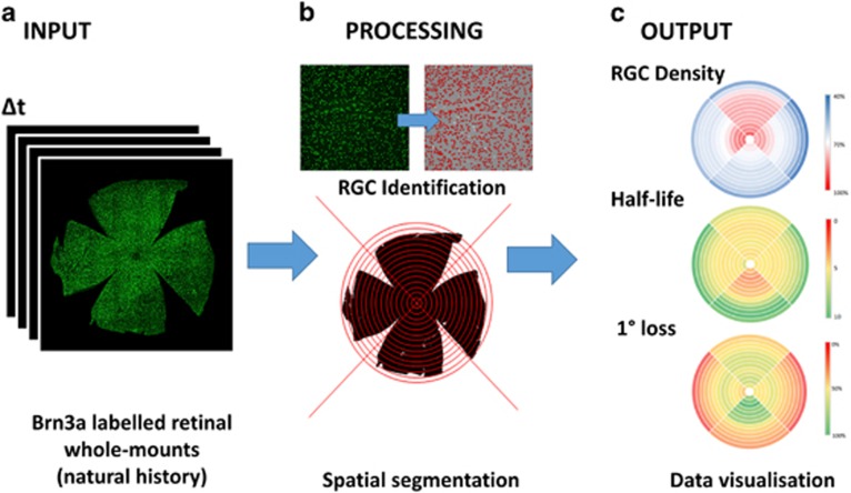
Figure 1.
Maximising the extraction of information from retinal whole-mounts. (a) Brn-3a retinal whole-mounts were obtained at different timeponts from established rodent models of optic neuropathy. (b) The algorithm we developed9 first segmented the RGC population from these whole-mounts before spatially segmenting the population into a series 60 of non-overlapping sectors defined relative to the position of the optic nerve head. (c) Assessment of longitudinal changes across the natural history of each model permitted the spatial-patterns in RGC density changes, half-life and the extent of primary degeneration to be evaluated and summarised as colourmaps

