Figure 1.
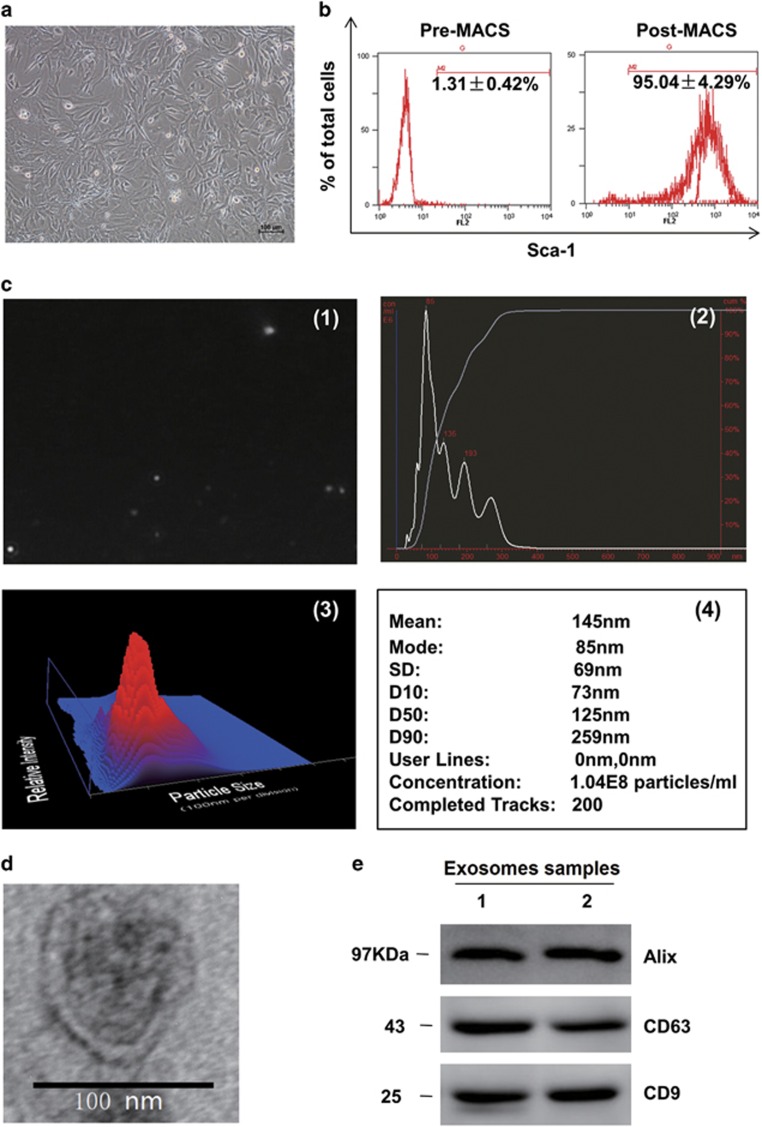
Characteristics of cardiac progenitor cells and exosomes. (a) The phase morphology of isolated CPCs growing on gelatin-coated dish; scale bar, 100 μm. (b) Flow Cytometry analyzed purified Sca-1+CPCs from the first preparation. Typical purity of isolation is >95% after magnetic beads sorting. (c) Nanoparticle trafficking analyzed the diameters and concentration of exosomes; 1 is a representative screen shot of the NTA videos, the bright white dot indicates one moving particle, (2) NTA estimated the size of the EVs between 90 and 300 nm, and the mode of these particles is 85 nm, and predict the proper concentration is around 1.04 × 10⁸ particles per ml, the dilution is 1 : 80, 3 is a heat map pattern of 2, and 4 is a detail statistical report. (d) Electron micrograph analyzed CPC-derived exosomes. The image showed small vesicles of approximately 100 nm in diameter. Scale bar, 100 nm. (e) Western blotting characterized CPC-exosomes. CPC-exosomes preparation was separated by SDS-polyacrylamide gel electrophoresis, and electroblotted to the poly vinylidene fluoride (PVDF) membrane, and probed with exosomes marker CD63, CD9, Alix

