Figure 1.
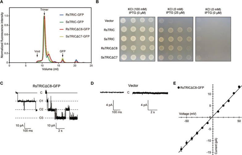
Functional characterization of prokaryotic TRIC proteins. (A) FSEC profiles for GFP-tagged RsTRIC (blue), SsTRIC (green), RsTRICΔC8 (red) and SsTRICΔC7 (yellow). The arrows indicate the elution positions of the void volume (void), the trimer of TRIC-GFP (trimer) and the free GFP (GFP). (B) Growth complementation assay of K+ auxotrophic strains harboring either the empty vector or the expression vectors encoding GFP-tagged prokaryotic TRIC proteins. A 10-fold dilution series was spotted on low-potassium medium plates. (C, D) Representative current traces recorded at −60 mV in a membrane patch of E. coli giant spheroplasts expressing GFP-tagged RsTRICΔC8 (n = 6) (C), or harboring the empty vector (n = 3) (D) in KCl-containing bath medium. In each panel, an enlarged trace is presented on the left. Uppercase letters, C and O1-O3, indicate closed and open states, respectively. Currents were recorded for 9 s in each step-pulse, and the single channel was observed as a triple event. (E) Current-voltage relationship (I-V curve), determined by measuring the current amplitude from −60 mV to +40 mV by 10 mV steps. The values presented represent mean ± SEM (n = 6).

