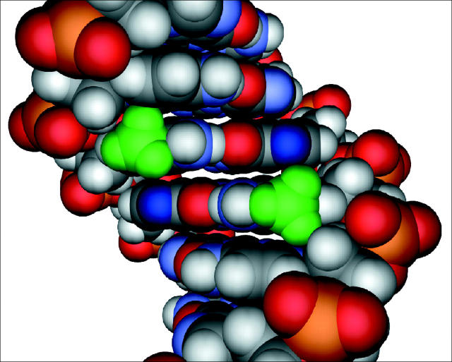
Figure 5.
Molecular model of the sequence 5′-GCmCGGC-3′. The methyl groups of 5mC are depicted in green, and the N7 position of guanines within the methyl-CpG dinucleotide are depicted in dark blue. Multiple sites within the methylated CpG dinucleotide are needed for strong binding by the MBD. All four major sites of contact, two methyl groups and two hydrogen bond accepting nitrogens, are in the major groove of the DNA within close proximity of one another. Disruption of MBD binding results from oxidative damage to any one of these four sites.

