
Figure 5.
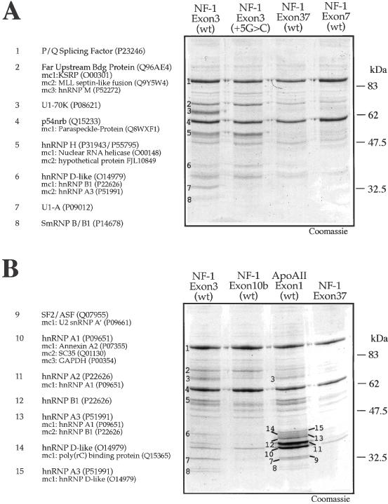
Ribonucleoprotein complexes in the 5′ss of NF-1 and Apo AII donor sites. (A) Coomassie Blue staining of a pulldown assay using beads coated with NF-1 exon 3 (wt), NF-1 exon 3 (+5G > C), NF-1 exon 7 (wt), and NF-1 exon 37 (wt) RNAs following incubation with HeLa nuclear extract. The numbers on the NF-1 exon 3 (wt) lane indicate the sequenced protein bands. The identity of each numbered band is shown to the left of the gel. (B) Coomassie Blue staining of a pulldown assay using beads derivatized with NF-1 exon 3 (wt), NF-1 exon 10b (wt) and Apo AII exon 1 (wt), and a control pulldown using exonic sequence from NF-1 exon 37 lacking a 5′ splice site. The numbers on the NF-1 exon 3 (wt) lane indicate the sequenced protein bands. On the left of the gel the identity of each band together with its Swiss-Prot Accession number is reported. Minor components (mc), when present, are also reported (in decreasing order of detection).