Figure 4.

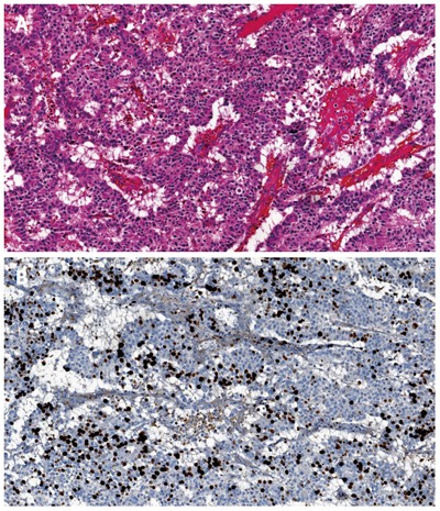
Morphological well differentiated neuroendocrine carcinoma of the pancreas (A, haematoxylin-eosin stain) and morphological well differentiated neuroendocrine carcinoma of the pancreas with Ki67 proliferative index of 30% (B).

Morphological well differentiated neuroendocrine carcinoma of the pancreas (A, haematoxylin-eosin stain) and morphological well differentiated neuroendocrine carcinoma of the pancreas with Ki67 proliferative index of 30% (B).