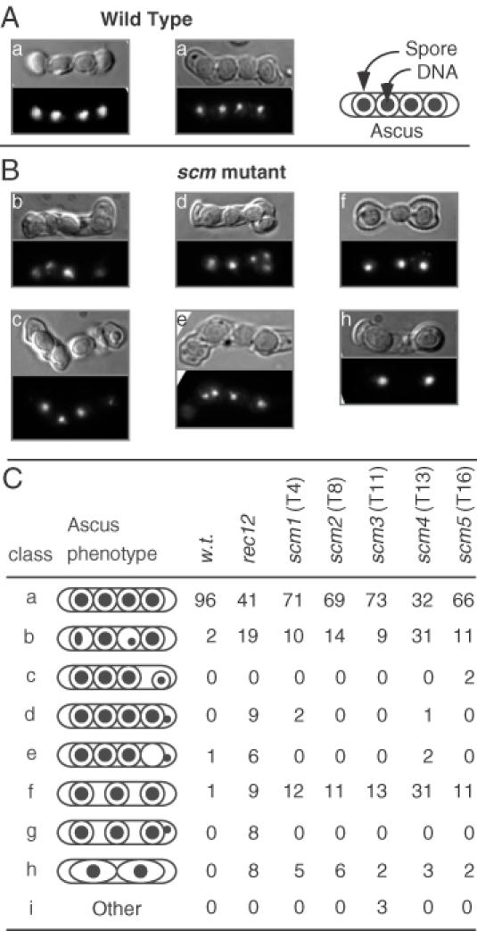
Figure 3.

Cytological analysis of meiotic chromosome segregation and ascus formation. Representative asci from wild-type (A) and scm mutant (B) strains. Differential interference contrast microscopy was used to analyze spore and ascus morphology (upper panel) and DAPI fluorescence microscopy revealed the distribution of DNA within each ascus (lower panel). Mutant phenotypes include unequal partitioning of chromosomes (b, c, e), failure of DNA to enclose within spores (d, e) and aberrant (c, d) or missing (f, h) spores. (C) Schematic representation of the ascus phenotypes. The outer oval depicts the ascus; the inner circles and ovals depict the spore walls; the filled circles depict the location and relative amount of DNA.
