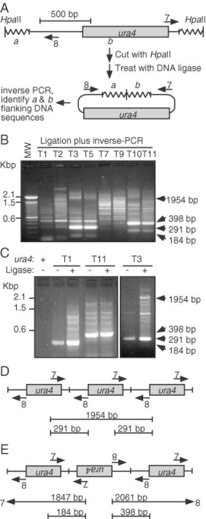
Figure 6.

Complex structures of ura4+ transgenes. (A) Expected transgene structure and inverse-PCR method to identify its location. Genomic DNA (wavy line) from each transformant was expected to harbor a single copy of the ura4+ transgene. In that case, inverse-PCR would produce a single DNA product that could be sequenced to identify the genomic locus into which the transgene had inserted. (B) Complex structure of transgenes. Genomic DNA was digested with HpaII, treated with DNA ligase, subjected to inverse-PCR using primers in panel ‘A’, and analyzed using agarose gel electrophoresis. (C) Analysis of junction fragments. Inverse-PCR was performed using samples with or without prior treatment with DNA ligase. Ligase-independent bands of characteristic size are diagnostic for different types of concatomeric fusions. (D) Schematic diagram of unit-length head-to-tail concatomers and diagnostic inverse-PCR product sizes. (E) Schematic diagram of head-to-head and tail-to-tail concatomers and diagnostic inverse-PCR product sizes. Product sizes such as those in (D) would also be generated.
