
Figure 1.
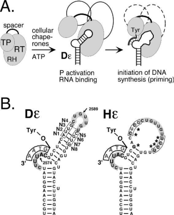
Hepadnavirus replication initiation. (A) Chaperone-dependent activation of P protein. P protein consists of TP, reverse transcriptase (RT) and RNaseH (RH) domains and a non-essential spacer. Cellular chaperones (symbolized by open ovals) are essential to enable P to bind its cognate ε RNA. A structural alteration in the upper stem allows a Tyr-residue in TP to act as acceptor for the 5′ terminal nucleotide of a short DNA oligonucleotide (thick line attached to Tyr) that is templated by the bulge region. This priming reaction can be reconstituted in RL. (B) Secondary structures of duck (DHBV) and heron HBV (HHBV) ε signals. Both Dε and Hε consist of a lower stem, a bulge and an upper stem, which is largely base paired in Dε but not in Hε. The conserved apical sequence, highlighted by grey background, is commonly referred to as loop although in Dε the 3′ terminal U is most probably base paired as shown; in Hε it is part of a large region lacking conventional base pairs due to various nucleotide exchanges (marked by asterisks). The nucleotide positions most sensitive to nucleases are indicated by arrows, and the randomized positions are marked N1 to N8. Equivalent Hε residues are boxed.