
Figure 6.
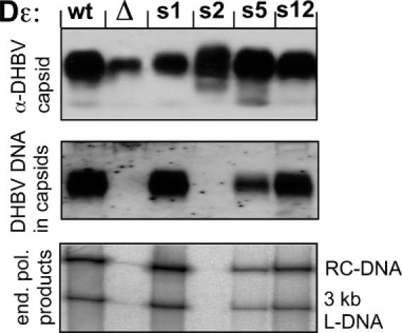
Functional analysis of selected aptamer sequences in a complete viral genome. LMH cells were transfected with DHBV expression plasmids carrying s1, s2, s5 and s12 derived Dε signals (s1, s2, s5 and s12), the wild-type genome (wt), or a modified wild-type genome lacking a functional ε signal (Δ). Cells were lysed and aliquots of the cytoplasmic lysate were run on a native agarose gel (upper two panels), blotted, and either probed with anti-DHBV capsid antibody (α-DHBV capsid) or hybridized with a DHBV specific DNA probe (DHBV DNA in capsids). Note that relative to the capsid signals, the DNA signals are increased in s1 and reduced in s5. Capsids immunoprecipitated from the lysates were subjected to an endogenous polymerase reaction; the products were separated by agarose gel electrophoresis and 32P-labelled viral genomes were visualized by phosphorimaging. The positions of the typical relaxed circular (RC) and linear (L) forms are indicated. The faint signal in lane Δ is due to contamination from the neighbouring lanes.