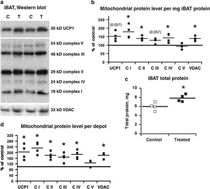
Figure 2.
Recruitment of BAT in wild-type mice treated with C12TPP for 16 days on a HFD at thermoneutrality. (a) Western blotting of mitochondrial proteins performed on interscapular BAT (iBAT) protein extract. (b) Mitochondrial protein content per mg tissue protein taken from quantification of western blotting as in a. (c) Total iBAT protein content. (d) Mitochondrial protein content per iBAT depot. In b–d, the mean and individual data points of four independent tissue extracts of each group are presented. For graphic presentation on b and d, the mean protein level of control iBAT was defined as 100% and the levels in iBAT from C12TPP-treated mice expressed relatively to this value. For statistics on b–d, the raw data were analyzed with Wilcoxon–Mann–Whitney test. Asterisks indicate significant differences between the control and C12TPP-treated groups.

