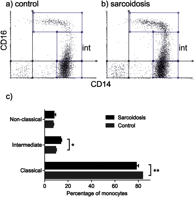
Figure 4. Intermediate CD14++ CD16+ blood monocytes are expanded in sarcoidosis.
Representative two colour flow cytometry dot pots from (a) control and (b) sarcoidosis subjects with intermediate monocytes indicated (int). Monocyte subsets were determined by extracellular antibody staining for CD14 and CD16. (c) Percentages of CD14++ CD16+ intermediate monocytes, CD14++ CD16− classical monocytes, and CD14+ CD16++ non-classical monocytes in patients with sarcoidosis (n = 14) and controls (n = 18). Results are presented as mean ± SEM; *p < 0.05, **p < 0.01 using two-way ANOVA with Sidak’s Post hoc test.

