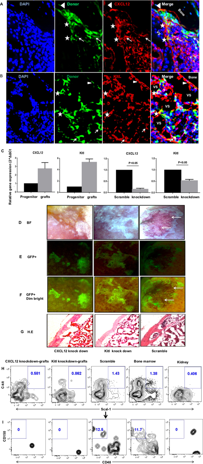
Figure 7. CXCL12 and Kitl play an important role in ectopic BM niche formation.

(A,B) Representative images of sections from GFP labeled CD133−CD55− progenitors derived KC grafts stained with antibody against CXCL12 (A) or Kitl (B). Cells expressing both GFP and CXCL12 (A) or GFP and Kitl (B) were indicated by triangles, stars and arrows. The triangles label cells located in endosteum. The stars label the cells in perivascular regions. The arrows label the cells in stromal regions. (C) Gene expression levels of CXCL12 and Kitl in freshly sorted CD133−CD55− progenitors and GFP+ donor derived cells harvested from KC grafts. (D) Knockdown efficiency of CXCL12 and Kitl was determined by qRT–PCR relative to scramble control. (E–H) Representative brightfield (E), GFP (F), GFP + dim brightfield (G) and H.E section (H) images of KC grafts derived from common progenitors transduced with lentivirus carried CXCL12 knockdown (left), Kitl knockdown (middle) or scramble control (right) constructs. Arrows indicate the vasculatures (only presented in the scramble control). (I,J) Representative FACS analysis of LSK (I; CD45+Lineage-c-Kit+Sca1+) and LT-HSC (J; LSKCD150+CD48−) frequency in KC grafts that were pre-gated for live, CD45+lineage− cells. The bone marrow and kidney cells were used as positive and negative controls, respectively.
