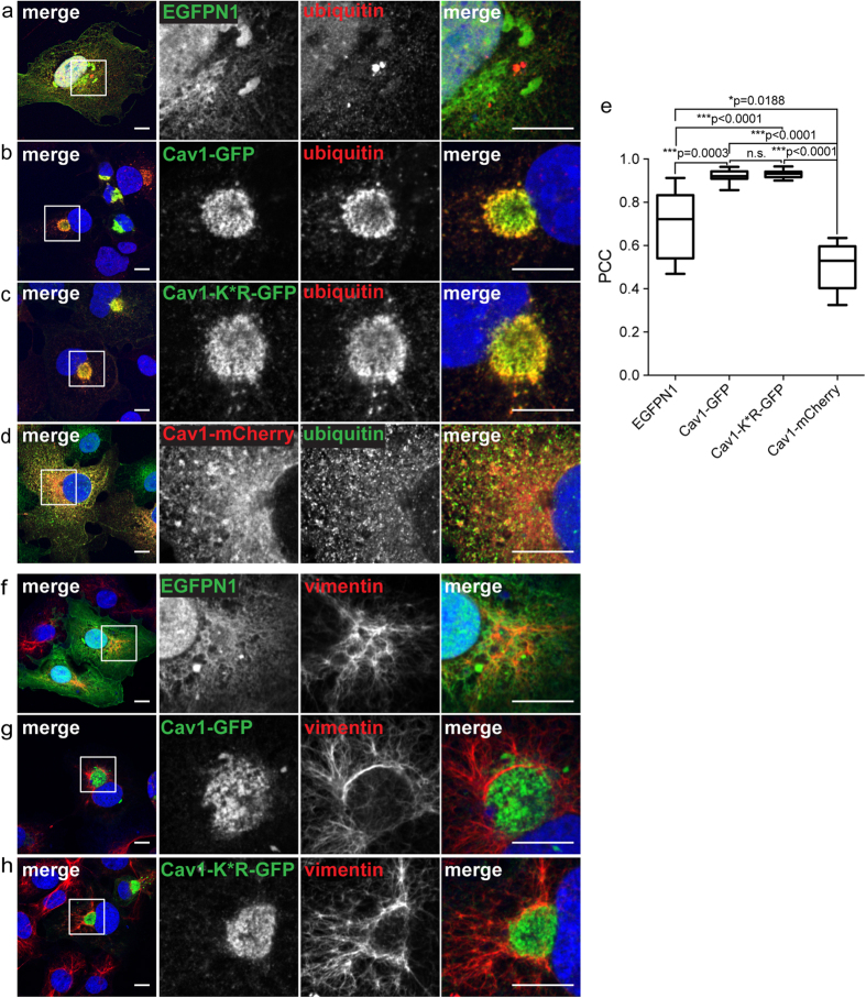
Figure 5. The perinuclear pool of Cav1-GFP is enriched in ubiquitinated proteins.
COS-7 cells transiently transfected with empty vector (EGFPN1), Cav1-GFP, Cav1-K*R-GFP or Cav1-mCherry were stained with either an anti-ubiquitin antibody (a–d) or chicken anti-vimentin antibody (f–h). In the merged images, GFP fluorescence is shown in green and antibody labeling is shown in red. For the Cav1-mCherry transfected cells, mCherry fluorescence is shown in red and antibody staining in green. DRAQ5 staining was used to label the nucleus (blue). White boxes indicate the areas of the zooms. Scale bars, 10 μm. (e) Quantification of colocalization of ubiquitin with the indicated constructs using Pearson’s correlation coefficient (PCC). n.s., not significant, P > 0.05; *P ≤ 0.05; **P ≤ 0.01, ***P ≤ 0.001; Mann-Whitney U-test. n = 9 cells per condition. Note that EGFP shows a high degree of colocalization with ubiquitin because both proteins are distributed throughout the cytoplasm and nucleus.

