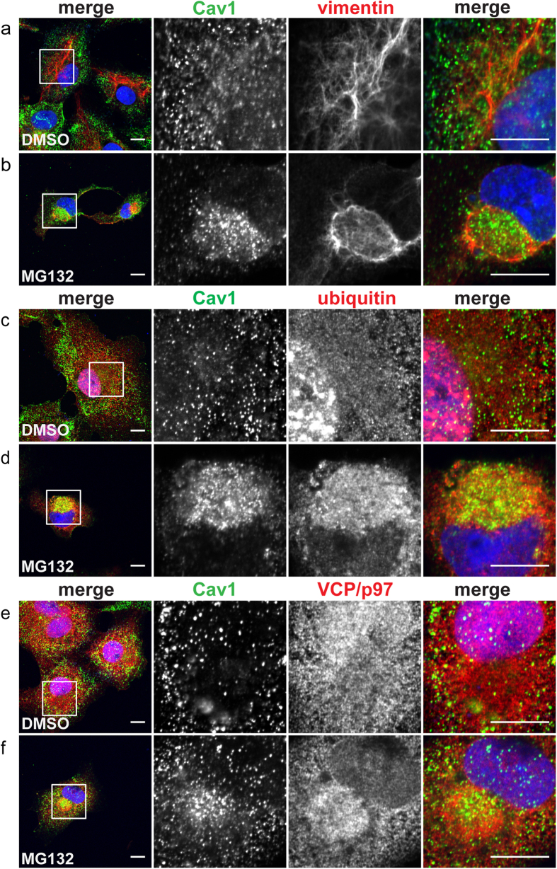
Figure 8. Endogenous Cav1 accumulates in aggresomes induced by proteotoxic stress.
COS-7 cells treated with either DMSO (a,c,e) or 5 μM MG132 (b,d,f) for 24 h were immunostained with rabbit anti-Cav1 polyclonal antibody and vimentin (a,b), ubiquitin (c,d), or VCP/p97 (e,f). Images were acquired using similar microscope settings for DMSO- and MG132-treated cells. Representative images from 2 independent experiments are shown. In the merged images Cav1 staining is shown in green and vimentin, ubiquitin or VCP/97 staining is depicted in red. DRAQ5 staining was used to label the nucleus (blue). White boxes indicate the areas of the zooms. Scale bars, 10 μm.

