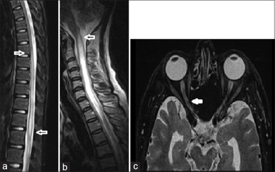
Figure 2.

Magnetic resonance imaging of the spinal cord and optic nerve in anti-myelin oligodendrocyte glycoprotein patients. (a) T2-weighted sagittal image of the cord in a 16-year-old male with anti-myelin oligodendrocyte glycoprotein and isolated longitudinally extensive transverse myelitis involving dorsal cord with extension to the conus (block arrow), (b) 42-year-old woman with anti-aquaporin-4 antibody and longitudinally extensive transverse myelitis in the cervical cord extending into the caudal brainstem (block arrow), (c) axial images of the orbit showing long segment hyperintense signals in the optic nerve (block arrow) and optic atrophy in the other in the same patient
