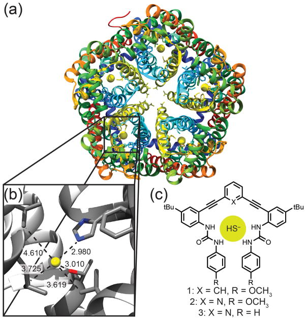
Figure 1.
(a) Protein structure of HSC (PDB:3TDX) showing five individual channels with the bound anion represented as a yellow sphere. (b) Enlargement of the binding pocket showing short contacts to His (2.980 Å), Thr (3.010 Å), Leu (3.725 Å) and Val (3.619 and 4.610 Å). Non-interacting helices are excluded for clarity. (c) Synthetic receptors 1–3.

