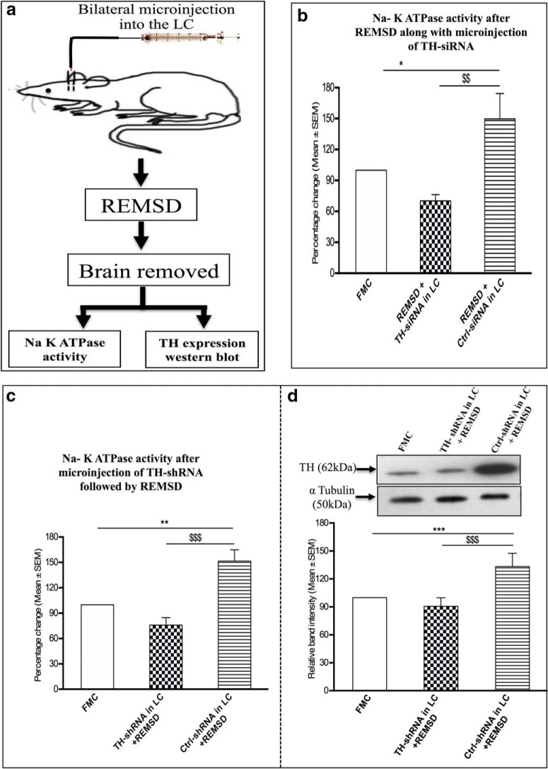
Figure 8.
a, Diagrammatic representation of protocol of experiment 3. b, Percentage (mean ± SEM) changes in Na-K ATPase activity in the brain homogenate of rats deprived of REMS after microinjection of TH-siRNA or Ctrl-siRNA bilaterally into the LC compared with the Na-K ATPase activity of FMC rats taken as 100%. c, Percentage (mean ± SEM) changes in Na-K ATPase activity in the brain homogenate of rats deprived of REMS after microinjection of TH-shRNA or Ctrl-shRNA bilaterally into the LC compared with Na-K ATPase activity in FMC rats taken as 100%. d, Percentage (mean ± SEM) change in TH protein expression in the brains of rats deprived of REMS for 96 h after the microinjection of TH-shRNA and Ctrl-shRNA bilaterally into the LC relative to FMC control taken as 100% are shown. *Compared with FMC; $compared with Ctrl-shRNA/Ctrl-siRNA in LC and subjected to REMSD. Significance level: ***p < 0.001; **p < 0.01; *p < 0.05: N = 5/group.

