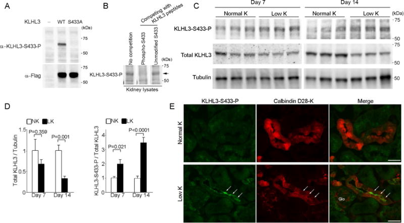
Figure 1. K+ depletion increases KLHL3 phosphorylation at S433 and decreases total KLHL3 levels.

(A) Western blot analysis of HEK cells expressing no KLHL3, Flag-tagged KLHL3WT or KLHL3S433A incubated with monoclonal anti-KLHL3S433-P antibody (upper panel) and anti-Flag antibody (lower panel). The signal detected by the anti-KLHL3S433-P is abolished by the S433A substitution. (B) Total kidney lysates were prepared from mice eating a normal diet. Western blots of the lysates were incubated with anti-KLHL3S433-P antibody without or with competition with immunizing KLHL3 phosphopeptide and non-phosphopeptide. Competition with the KLHL3 phosphopeptide but not the non-phosphopeptide eliminates the antibody signal (arrow), confirming the specificity. (C) KLHL3S433-P and total KLHL3 levels in the kidneys of wild-type mice fed a normal K+ diet (NK) or a low-K+ diet (LK) determined by Western blot analysis in biological replicates. (D) Quantitation of total KLHL3 and KLHL3S433-P levels in the kidney described in (C) (n = 6 or 7 for day 7 and n = 5 for day 14). Data are expressed as means ± SEM. (E) Kidney sections stained for α-KLHL3S433-P (green, indicated by arrows) and α-calbindin D28-K (a marker for distal convoluted tubule cells, red) in mice eating a normal-K+ or a low-K+ diet. KLHL3S433-P is increased at the apical membrane of distal convoluted cells (which express NCC). Scale bars represent 50 μm. Glo, glomeruli.
