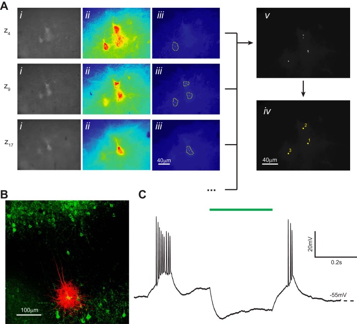
Fig. 10.
Computer vision-aided automatic cell detection of ArchT-EYFP-positive cells. A: computer vision processing of images acquired with epifluorescence optics detects fluorescent neurons and identifies their x, y, z coordinates. Three representative z sections are shown from a complete experiment (20 total z sections) using brain slices prepared from a mouse injected with HSV-ArchT-EYFP virus. i, Original image after histogram equalization; ii, pseudo-colored image after thresholding; iii, superimposed cell-like contours detected after a series of varying thresholds; iv, centroids of detected contours are accumulated from z sections; v, centroids from the complete z scan (20 z sections) are clustered and the final coordinates calculated. B: representative ArchT-EYFP-positive cell (green) filled with Alexa Fluor 568, postfixed, and immunolabeled with the anti-GFP antibody. Images are acquired using confocal microscopy. C: representative current-clamp recording trace of a layer 5 bursting cell hyperpolarized in response to light (550 nm) activation (green bar).

