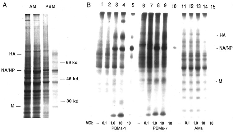
Figure 3.
A, Autoradiograms examining synthesis of influenza A virus (IAV) proteins by alveolar macrophages (AMs; lanes 1 and 2) and autologous peripheral blood–derived monocytes-macrophages (PBMs; lanes 3 and 4). Cells were sham-exposed (lanes 1 and 3) or exposed (lanes 2 and 4) to IAV, pulse labeled 4–6 hours after exposure and analyzed with polyacrylamide gel electrophoresis. B, Viral protein synthesis by PBMs cultured for 1 day (PBMs-1; lanes 1–5), PBMs cultured for 7 days (PBMs-7; lanes 6–10), and AMs (lanes 11–15) exposed in vitro to influenza A/AA/Marton/43 H1N1 at a multiplicity of infection (MOI) of 0 (sham-exposed cells; lanes 1, 6, and 11), 0.1 (lanes 2, 7, and 12), 1.0 (lanes 3, 8, and 13), and 10 (lanes 4, 9, and 14). Lanes 5, 10, and 15 represent immunoprecipitation of lysates of virus-exposed (MOI, 10) PBMs-1, PBMs-7, and AMs respectively, using goat polyclonal antiserum directed against subtype-specific hemagglutinin (HA), neuraminidase (NA; which comigrates with nucleoprotein [NP]), matrix protein (M), and murine monoclonal antibody to NP. Lysates were derived from equal numbers of cells pulsed 4–6 hours after exposure to the virus.

