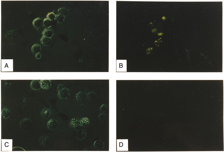
Figure 6.
Photomicrographs of alveolar macrophages (AMs) exposed to fluorescein isothiocyanate (FITC)–labeled influenza A virus (IAV). A, C, Light photomicrographs. B, D, Corresponding fluorescence detection. Left panels are light images with reduced lighting to diminish potential quenching of fluorescence in the field. A, B, AMs exposed to the virus in the presence of anti-FITC antibody. C, D, AMs exposed to virus in the presence of the control anti–yellow fever virus antibody. Cells are shown before addition of ethidium bromide, and the results indicate binding and/or internalization of the virus. Addition of ethidium bromide did not detectably alter the findings, suggesting that the virus had internalized.

