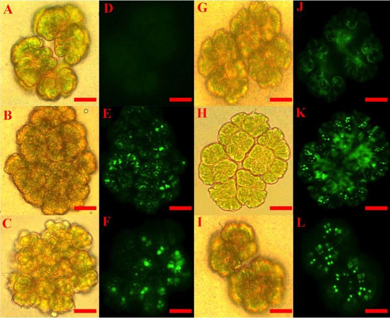
Figure 1. Staining of ROS positive colonies of Botryococcus braunii.
CellROX dye was used to detect ROS in vivo by observing fluorescent B. braunii colonies after 60 min treatments. B. braunii colonies under white light (A, B, C, G, H and I) and fluorescent excitation (D, E, F, J, K and L). Control without treatment (A, D); and treatments with 100 mM NaCl (B, E), 120 mM NaHCO3 (C, F), 3 mM SA (G, J), 10 µM MeJA (H, K), and 4.33 mM acetic acid (I, L).

