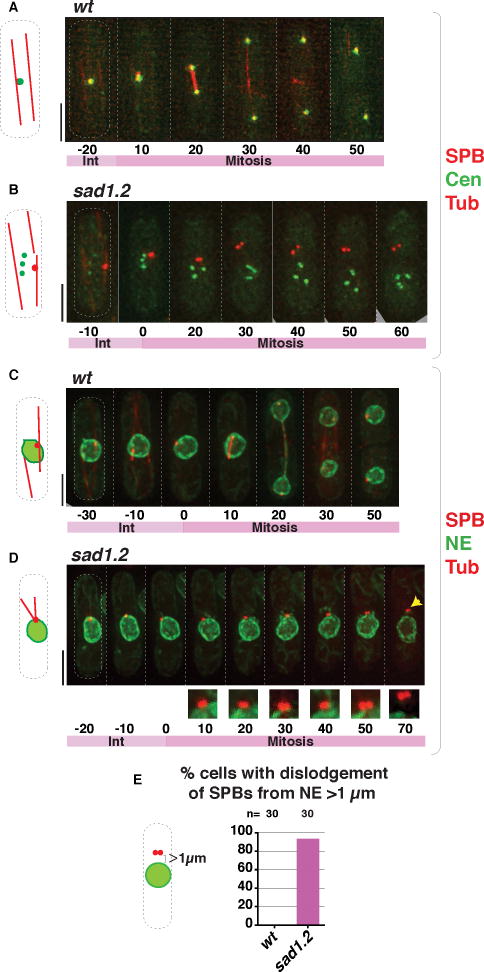
Figure 4. Failed SPB insertion in mitotic sad1.2 cells leads to abolition of spindle formation.
(A–B) Frames from films of proliferating cells at 36 °C; SPBs seen via Sid4-mCherry, centromeres via Ndc80-GFP (Cen) and spindles via ectopically expressed mCherry-Atb2 (nda3 promoter controlled). Growth for 4 h at 36 °C led to complete centromere-LINC dissociation in the sad1.2 setting (B); SPB separation defects ensued. (C–D) Visualization of the NE via Ish1-GFP, cells grown as in (A–B). As in bqt1Δ meiosis (Figure 1G), the duplicated SPBs detach from NE during sad1.2 mitosis. Numbers indicate mitotic progression in minutes; t=0 is just before SPB duplication. (E) Quantitation of phenotypes shown in (D) from six independent experiments. Dislodgement of the SPB from the NE is never observed in wt cells. n is the total number of cells scored. See also Figure S5.

