
Figure 2.
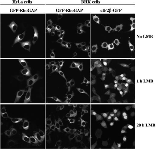
In either stably transduced HeLa or BHK cells, GFP-p50RhoGAP shows a cytoplasmic staining, with some enrichment at the Golgi. Inactivation of CRM1 by LMB (5 ng/ml) did not change this localisation, even after 20 h of treatment. In contrast, already 1 h of LMB treatment shifted the CRM1 cargo eIF2β-GFP from exclusively cytoplasmic to mainly nuclear. Images of the paraformaldehyde-fixed samples were taken by confocal laser scanning microscopy.