
Figure 1.
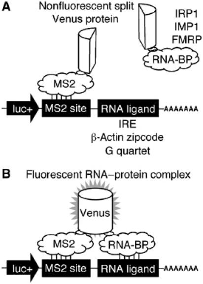
A TriFC method to study RNA–protein interactions in living cells. A portion of the Venus fluorescent protein is attached to a reporter mRNA by the bacteriophage MS2 coat protein–RNA interaction. The complementing portion of Venus is fused to an RNA-binding protein (A). If the RNA-binding protein interacts with a sequence of interest within the reporter mRNA, the two portions of Venus are brought into close proximity to form a fluorescent product (B).