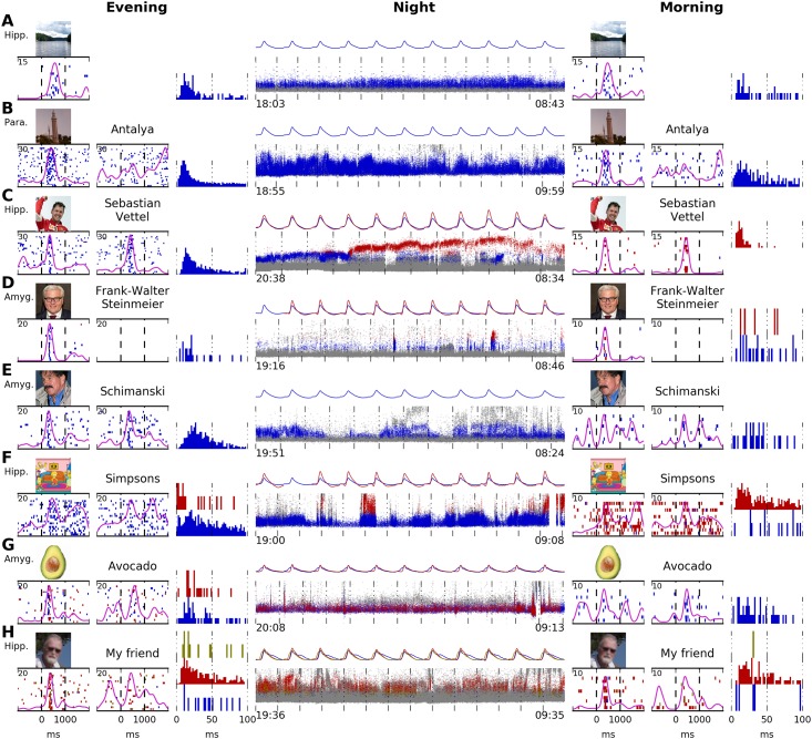
Fig 10. Tracking of selectively responding neurons over an entire night.
A–H show data from eight patients. Continuous unit recordings started in the evening and ended the next morning. “Screening sessions” were performed at the beginning and in the end of each recording. Displayed are raster plots for one stimulus image per screening session. Inter-stimulus interval histograms for the evening and morning are displayed. In all patients but A, written names corresponding to the images were also presented. The middle column (“Night”) shows the activity of units tracked automatically during the entire recording. Each small dot marks the time point and maximal voltage of one action potential. Colors correspond to the raster plots from the screening sessions: units marked in gray do not respond to the images/written names. Units marked in blue, red, or yellow respond to the images/written names as shown in the raster plots. Mean waveforms of all responsive units are displayed for each hour recorded. A Stable waveform and response pattern. B Amplitude variations are visible. As typical for parahippocampal units, unit does not respond to the written name. C An amplitude shift in the responsive neuron (possibly caused by micro-movement of the electrode) results in the detection of two different units, most likely belonging to one neuron. D Two responsive clusters are generated. No response to the written name. E Stable waveform, but very weak response in the morning. F Solid response in the evening and morning, but with separate units. No definite conclusion about the success of tracking can be made. G The blue cluster generates most of the response. The red cluster also contributes to the response. Both clusters are tracked with a stable waveform. H Similar to G, with three responsive clusters. The red cluster generates most of the response. The blue and yellow clusters contribute to the response. All three clusters have a stable waveform. Hipp., hippocampus; Para., parahippocampal cortex; Amyg., amygdala. Stimulus pictures displayed here have been replaced by similar pictures for legal and privacy reasons. Copyright notes: A “Lake” by J. Niediek is licensed under CC BY 4.0 B “Antalya” by J. Schmidtkunz is licensed under CC BY 4.0 C cropped from “[…] Sebastian Vettel (Ferrari)” by Morio, CC BY-SA 4.0, Wikimedia Commons (https://commons.wikimedia.org/wiki/File:Sebastian_Vettel_2015_Malaysia_podium_2.jpg D cropped from “50th Munich Security Conference 2014: Vitali Klychko and Frank-Walter Steinmeier […]” by Mueller / MSC (Marc Müller), CC BY 3.0 DE, Wikimedia Commons (https://commons.wikimedia.org/wiki/File:MSC_2014_Klychko-Steinmeier3_Mueller_MSC2014.jpg) E “[…] Horst Schimanski […]” by H. Schrapers is licensed under CC BY-SA 2.5, Wikimedia Commons (https://commons.wikimedia.org/wiki/File:HorstSchimanski.jpg) F cropped from “Simpsons 20 Years” by Gabriel Shepard, CC BY-SA 3.0, DeviantArt (http://gabrielshepard.deviantart.com/art/Simpsons-20-Years-124858104) G “Avocado” by D. E. Bruschi is licensed under CC BY 4.0 H “My friend” by J. Niediek is licensed under CC BY 4.0.

