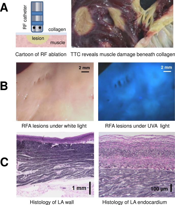
Fig 2. Endocardial collagen layer masks RF-induced damage to atrial muscle below.
A. Left: a cartoon of RF catheter ablating atrial endocardial surface. Right: an example of ablated human left atrium after TTC-staining. By peeling off the collagen layer, RF damage to the muscle below can be readily seen (ablated tissue shows as pale areas devoid of red triphenylformazan dye). B. Unstained endocardial surface of human left atrium with multiple RF lesions under either room light or UV illumination. Note the limited contrast between lesion sites and unablated, healthy tissue. C. Histology of left atrial wall shows layers of atrial muscle sandwiched between endocardial collagen layer and epicardial fat. A close-up of endocardial layers reveals interwoven fibers of collagen (pink) and elastin (black).

