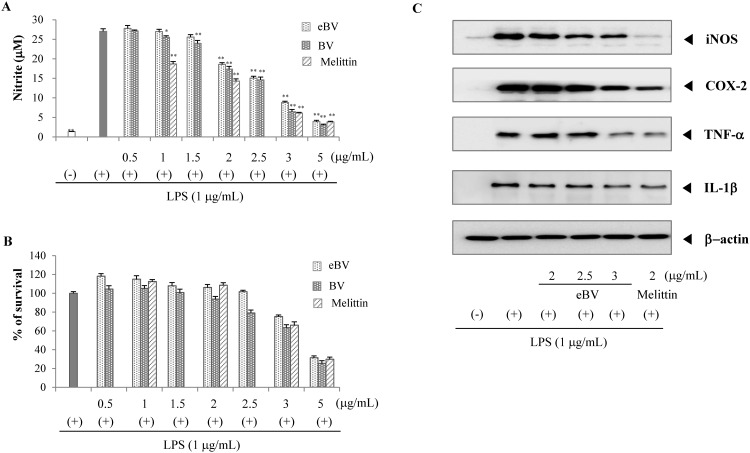
Fig 4. Effect of eBV on LPS-induced NO production and protein expression in macrophage cells.
(A) RAW 264.7 cells were stimulated with LPS (1 μg/mL) in the presence or absence of eBV. After 20 h, the cultured media were collected, and nitrite concentration was analyzed using Griess reaction. The data are expressed as mean ± SD of triplicate tests. *p < 0.05 and **p < 0.01 indicate statistically significant difference compared with the LPS (+) group. (B) Cell viability was measured by the MTT method as described in the Materials and Methods. (C) The effect of eBV on iNOS, COX-2, TNF-α, and IL-1β protein expression in LPS-stimulated RAW 264.7 cells. RAW 264.7 cells (5 × 10⁵ cells/mL) were incubated for 24 h and then treated with LPS (1 μg/mL) and eBV for an additional 4 h and 20 h. After incubation, total cell extracts were obtained and subjected to Western blot analysis as described in the Materials and Methods. The data are representative of three separate experiments. β-actin was used as an internal standard.

