
Figure 2.
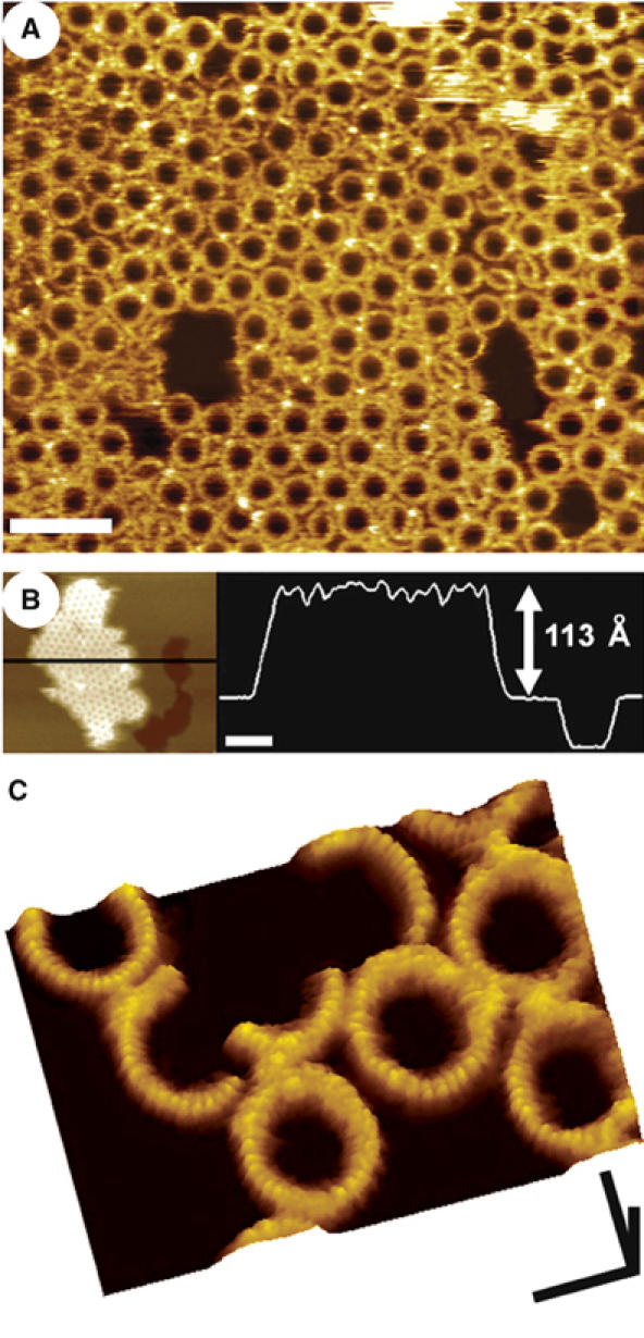
AFM images of the PFO prepore complex associated with supported lipid bilayers containing cholesterol. (A) The prepore-trapped PFOY181C complexes form a largely uniform population of ring structures, 38 nm in outer diameter. Scale bar: 100 nm. (B) These complexes measure 113±5 Å high from the top of the membrane surface, similar to the height of the water-soluble monomer (Figure 1). The darkest region in this figure is a large defect in the membrane, where the tip is directly in touch with the mica substrate. Scale bar: 100 nm. (C) At smaller scan sizes, higher resolution features are clearly discerned, including the 25 Å periodic arrangement of subunits in the complex and a slight 7 Å decrease in height of each subunit from its outermost to its innermost edge. Scale bars: x, y, 25 nm; z, 10 nm.